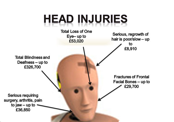
Head injury refers to trauma to the head. This may or may not include injury to the brain. However, the terms traumatic brain injury and head injury are often used interchangeably in the medical literature.Head trauma has a poor potential for good outcome.Deaths from head trauma occurs at three time points after the injury.,immediately after the injury,2 hours after the injury,and approximately 3 weeks after the injury.
INCIDENCE
The incidence (number of new cases) of head injury is 300 per 100,000 per year (0.3% of the population), with a mortality of 25 per 100,000 in North America and 9 per 100,000 in Britain. Head trauma is a common cause of childhood hospitalization.
CAUSES
Common causes of head injury are motor vehicle traffic collisions, home and occupational accidents, falls, and assaults. Bicycle accidents are also a common cause of head injury-related death and disability, especially among children.
Types of head injury:
Scalp lacerations, Skull fractures, Minor head trauma, Major head trauma,
Scalp lacerations
Scalp lacerations are the most minor type of head trauma. Because the scalp contains many blood vessels with poor constrictive abilities,most scalp injuries are associated with profuse bleedingThe major complication associated with scalp laceration is infection.
Skull fracture
Skull fractures arefrquently with head traumaThere are several types of skull fractures
Linear
Depressed
Simple,communited,or compound fracture
Basillar skull fracture
CLINICAL MANIFESTAIONS OF DIFFERENT TYPES OF SKULL FRACTURE
Frontal fracture-CSF rhinorrhea ,pneumocranium
Orbital Fracture-Periorbiatal ecchymosis,(racoon eyes)
Temporal Fracture-Oval shape bruise behind the ear in the mastoid region(Battle s sign)CSF Otorrhea
Parietal Fracture-Deafness ,CSF or brain otorrhea,bulging of the tympanic membrane caused by blood or CSF,facial paralysis,loss of taste,Battle s sign
Basillar skull fractures-CSF or brain otorrhea,bulging of the tympanic membrane caused by blood or CSF,Battles sign,tinnitus or hearing difficulty,facial paralysis,vertigo.
Common Symptoms of skull fracture can include:
leaking cerebrospinal fluid (a clear fluid drainage from nose, mouth or ear) may be and is strongly indicative of basilar skull fracture and the tearing of sheaths surrounding the brain, which can lead to secondary brain infection.
visible deformity or depression in the head or face; for example a sunken eye can indicate a maxillar fracture
an eye that cannot move or is deviated to one side can indicate that a broken facial bone is pinching a nerve that innervates eye muscles
wounds or bruises on the scalp or face.
Basilar skull fractures, those that occur at the base of the skull, are associated with Battle’s sign, a subcutaneous bleed over the mastoid, hemotympanum, and cerebrospinal fluid rhinorrhea and otorrhea.
MINOR HEAD TRAUMA
Brain injuries are classified as major and minor.Concussion is a minor head injury.It is a sudden transient mechanical head injury with distruption of the neural activity and loss of consciousness.The patient maynot loss full consciousness with the head injury
Signs of concussion includes a brief distruption of LOC,amnesia regarding the event and headacheThe manifestations are short term duration.If the patient has not loss consciousness or if the loss of consciousness lasts for more than 5minutes,the patient is discharged from the with notification to return back if the symptoms persists.
Post concussion syndrome is usually seen from 2weeks to 2 months after the concussion.Symptoms includes persistent headache,lethargy,personality and behavioural changes,short attention span,decreased short term memory,and changes in intellectual ability.This affects the patients ability for activities of daily living
MAJOR HEAD TRAUMA
Major head trauma includes cerebral contusions and lacerations.Both injuries represent severe trauma to the brain..Contusions and intercerebral lacerations are generally associated with closed injuries
A contusion is the bruising of the brain tissue within a focal area that maintains the integrity of the piamater and arachnoid layers.A contusion develops areas of hemorrhage,infarction,necrosis and edema.With contusion the phenomenon of counter coup injury is often noted.Damage from coup counter coup injury occurs occurs due to the mass movement of brain inside the skull.Contusions or lacerations occurs at the site of direct impact on the skull(coup)and a secondary area on the opposite side away from the injury(countercoup)leading to multiple contused areas.Bleeding around the contusion site is usually minimal,and the blood is reabsorbed slowly.Neurological findings demonstrates generalized disturbance in LOC.Seizures are a common complication of brain contusion.
Laceration involves actual tearing of barin tissue and often occurs in association with depressed and compound fractures and penetrating injuries.Tissue damage is severe but surgical repair cannot be done because of the texture of the brain tissue.If bleeding is deep into the brain parenchyma then focal neurological symptoms are suspected.
When the head injury is major hemorrhage is suspected.This hemorrhage manifests as a space occupying lesion which is accompanied by unconsciousness,hemiplegia on the contralateral side,and a dilated pupil.
As hematoma expands,symptoms of increased ICP becomes more severe,prognosis becomes more severe because of intercerbral hemorrhage.
PATHOPHYSIOLOGY
1. Diffuse axonal injury is a widespread axonal damageoccuring after a mild,moderate or severe injury
2. Due to the etiological factors(immediate tearing of the axon from the traumatic injury)
3. The damage occurs primarily around the axons in the subcortical white matter of the cerebral hemispheres,basal ganglia,thalamus and the brain stem
4. The axons will get sheared Axonal disconnection and axonal swelling occurs
COMPLICATIONS
Epidural hematoma
Subdural hematoma
Intracerebral hematoma
Subarachnoid hemorrhage
CLINICAL FEATUERES
Presentation varies according to the injury. Some patients with head trauma stabilize and other patients deteriorate. A patient may present with or without neurologic deficit.
Patients with concussion may have a history of seconds to minutes unconsciousness, then normal arousal. Disturbance of vision and equilibrium may also occur.
Common symptoms of head injury include coma, confusion, drowsiness, personality change, seizures, nausea and vomiting, headache and a lucid interval, during which a patient appears conscious only to deteriorate later.[5]
Because brain injuries can be life threatening, even people with apparently slight injuries, with no noticeable signs or complaints, require close observation. The caretakers of those patients with mild trauma who are released from the hospital are frequently advised to rouse the patient several times during the next 12 to 24 hours to assess for worsening symptoms.
The Glasgow Coma Scale is a tool for measuring degree of unconsciousness and is thus a useful tool for determining severity of injury. The Pediatric Glasgow Coma Scale is used in young children.
Diagnosis and prognosis
Head injury may be associated with a neck injury. Bruises on the back or neck, neck pain, or pain radiating to the arms are signs of cervical spine injury and merit spinal immobilization by application of a cervical collar and possibly a long board
If the neurological examination is normal this is reassuring, however a serious intra-cranial injury may still be present. If the patient developing a worsening headache, has a seizure, develops one sided weakness, or has persistent vomiting than the patient should be advised CTimmediately
Imaging
The need for imaging in patients who have suffered a minor head injury is debated. A non-contrast CT of the head should be performed immediately in all those who have suffered a moderate or severe head injury.
MANAGEMENT OF HEAD-INJURED PATIENTS
The management of head-injured patients depends on the GCS following resuscitation.
Patients with a mild head injury (GCS 14-15) should be admitted to a ward where thorough and frequent neurological observations can be ensured.
CT scan of the patient’s head should be performed promptly, and the local neuro-surgical unit contacted.
Patients with a mild head injury should be observed until they make a complete neurological recovery and are only discharged if a responsible adult can supervise them at home for a further few days.
All patients with a GCS of 13 or less should receive a CT scan of their head although many authorities would advocate a CT scan on all whose GCS is not normal.
Those with an acute lesion on CT scan or evidence of diffuse cerebral oedema should be urgently discussed with the local neurosurgical unit, with the CT images transferred immediately, either by computer image-link or courier.
All CT scans should be accompanied by a provisional radiology report from the referring hospital.
Other indications for neurosurgical referral include compound depressed skull fracture, severely depressed fracture, deteriorating GCS score even with a normal scan and cerebrospinal fluid otorrhoea and rhinorrhoea.
The following details are necessary when making a neurosurgical referral: name, age, sex, date, time and mechanism of injury, initial GCS on scene (documented by paramedics) and GCS following resuscitation (before administration of anaesthetic agents should they be required), evidence of deteriorating GCS, pupil reaction, vital observations, previous medical and drug history, previous functional ability and mobility in the case of elderly patients, other injuries and management of the patient since injury.
INITIAL MANAGEMENT
The fundamental goals of resuscitation of the head-injured patient are the restoration of circulating volume, blood pressure, oxygenation, and ventilation.
The physician should initiate maneuvers that serve to lower ICP and do not interfere with these aims as early as possible during resuscitation of any patient with a head injury.
Treatment modalities such as hyperventilation and mannitol administration that have the potential of exacerbating intracranial ischemia or interfering with resuscitation should be reserved for patients who show signs of intracranial hypertension such as evidence of herniation or neurological deterioration.
USE OF MANNITOL:
Mannitol is effective for control of raised ICP after severe TBI. Effective doses range from 0.25 to 1 g/kg/body weight.
USE OF BARBITURATES IN THE CONTROL OF INTRACRANIAL HYPERTENSION
High-dose barbiturate therapy is efficacious in lowering ICP and decreasing mortality in the setting of uncontrollable ICP refractory to all other conventional medical and surgical ICP-lowering treatments, in salvageable TBI patients. Utilization of barbiturates for the prophylactic treatment of ICP is not indicated. The potential complications attendant on this form of therapy mandate that its use be limited to critical care providers and that appropriate systemic monitoring be undertaken to avoid or treat any hemodynamic instability. When barbiturate coma is utilized, consideration should also be given to monitoring arteriovenous oxygen saturation as some patients treated in this fashion may develop oligemic cerebral hypoxia.


